How to Interpret a Thyroid Panel: Why It's the Last Place You Should Start
Mar 16, 2026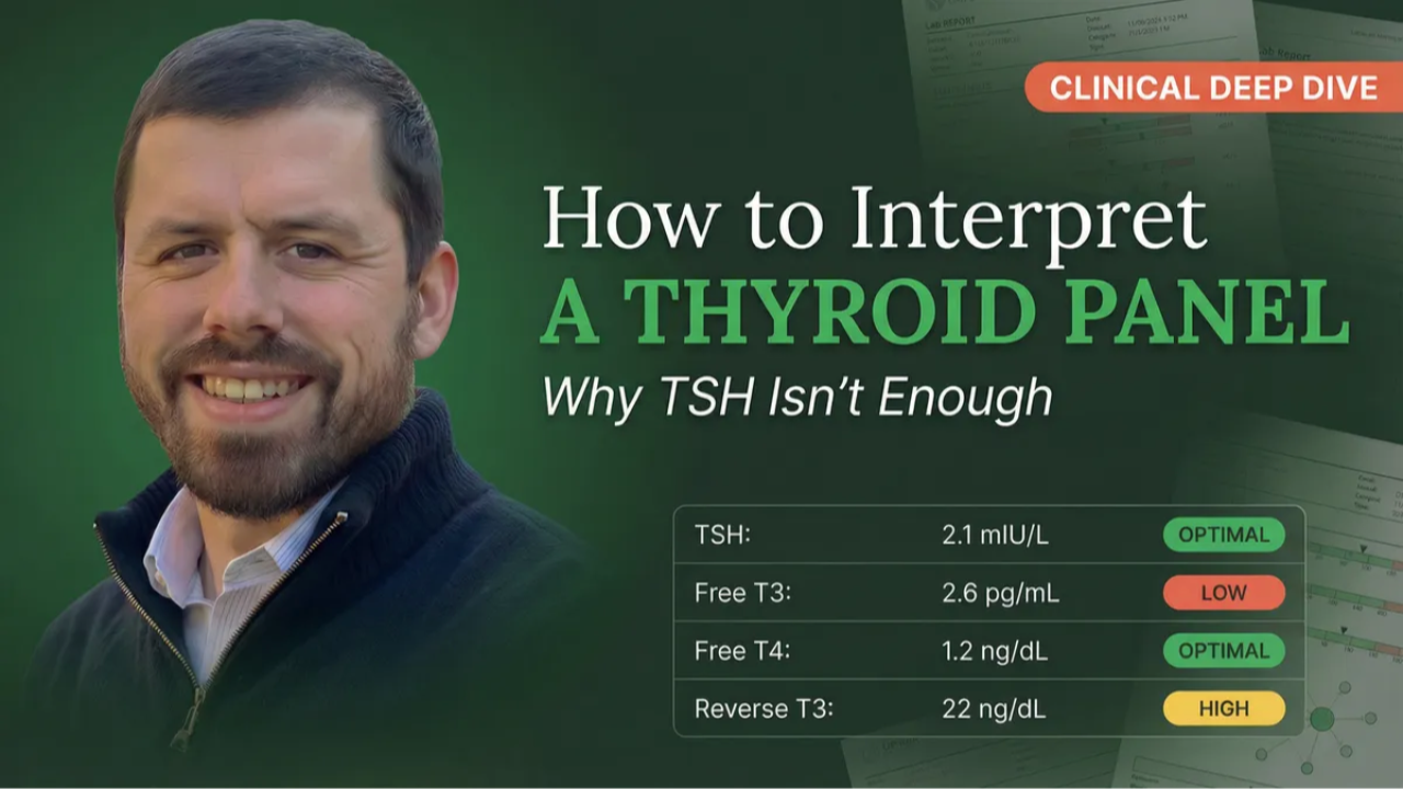
Clinical Deep Dive
How to Interpret a Thyroid Panel: Why It’s the Last Place You Should Start
The Full Panel, Three Clinical Patterns & Why Blood Sugar Comes First
Every client wants to start with thyroid. “I think it’s my thyroid.” And they might be right — but the thyroid doesn’t break in isolation. It is the canary in the coal mine for systemic health, reflecting dysfunction in blood sugar, liver function, adrenal stress, chronic inflammation, iron status, and nutrient cofactors. If you jump straight to thyroid protocols without addressing the foundation, you will fail.
This guide covers why thyroid assessment comes last in the clinical hierarchy (not first), the complete panel with functional optimal ranges, three primary clinical patterns that account for the vast majority of what you’ll see in practice, why antibodies are a layer and not a pattern, and the conversion problem that conventional testing misses entirely.
Twelve percent of the US population will develop thyroid conditions during their lifetime, with 20 million Americans currently affected. Sixty percent of those with thyroid disease remain unaware of their condition. Women face 5–8 times higher risk than men.1 And those statistics reflect only overt disease — including subclinical dysfunction would make the numbers significantly worse.
Why Thyroid Assessment Comes Last, Not First
In the Three-Tier Decision Tree, thyroid sits downstream of blood sugar, liver, adrenals, inflammation, iron, and key nutrient cofactors. This isn’t arbitrary. It reflects the physiological reality that thyroid dysfunction is almost always driven by upstream imbalances:
Everything Connects to the Thyroid
→ Blood sugar dysregulation suppresses thyroid conversion — insulin resistance impairs the enzymes that convert T4 to T3
→ Liver dysfunction impairs T4 → T3 conversion — 60% of conversion happens in the liver
→ Adrenal stress shunts T4 → Reverse T3 instead of active T3 — cortisol is the #1 conversion blocker
→ Chronic inflammation suppresses thyroid function at every level
→ Iron deficiency impairs thyroid hormone production — ferritin below 50 ng/mL impairs conversion2
→ Selenium, zinc, magnesium, and vitamin D are all required for thyroid function3
“You can’t thyroid your way out of a blood sugar problem.” Fix the metabolic foundation first. Then assess what’s left. 80% of the initial strategy is addressing the foundation — blood sugar, gut, stress, nutrients. 20% is direct thyroid support. This ratio surprises practitioners, but it’s what works.
The Complete Thyroid Panel
Six markers, three categories: the pituitary signal (TSH), the hormones (Free T4, Free T3, Reverse T3), and the autoimmune flag (TPO, TgAb). Every thyroid assessment starts here.
TSH — The Pituitary Signal
Conventional: 0.5–4.5 mIU/L
Functional Optimal: 1.0–2.5 mIU/L
TSH is produced by the pituitary, not the thyroid — it’s a demand signal, not a function marker. When thyroid hormones are low, TSH rises (“make more!”). When they’re high, TSH drops. TSH is the LAST marker to change. Symptoms and Free T3/T4 shift before TSH does. A “normal” TSH does not rule out thyroid dysfunction — it can be “normal” in secondary hypothyroidism, early Hashimoto’s, and stress-induced suppression.4
Free T4 — The Raw Material
Conventional: 0.8–1.8 ng/dL
Functional Optimal: 1.1–1.6 ng/dL
T4 is the raw material that gets converted to either Free T3 (active) or Reverse T3 (inactive). Normal Free T4 + low Free T3 = the conversion step is broken, not the gland. This distinction changes the entire intervention approach.
Free T3 — The Active Hormone
Conventional: 2.3–4.2 pg/mL
Functional Optimal: 3.0–4.0 pg/mL
Free T3 is the most metabolically active thyroid hormone — this is what your cells actually use. It correlates best with symptoms, more than TSH, more than Free T4. If you could only order one thyroid hormone marker, this would be it. Low Free T3 with normal Free T4 = conversion problem. Low Free T3 with low Free T4 = production problem.5
Reverse T3 — The Stress Brake
Conventional: 8–25 ng/dL
Functional Optimal: 10–17 ng/dL
The body produces Reverse T3 instead of Free T3 when it wants to slow down metabolism — the emergency brake. When you see low Free T3, Reverse T3 tells you why: elevated rT3 means stress/inflammation is shunting conversion. Low rT3 with low T3 means nutrient deficiency — the enzymes lack cofactors to convert T4 in either direction. This distinction changes the entire intervention strategy.6
TPO & Thyroglobulin Antibodies — The Autoimmune Flag
Functional: TPO <10 IU/mL • TgAb <1 IU/mL
Conventional: TPO <35 IU/mL • TgAb <20 IU/mL
TPO antibodies attack the enzyme that produces thyroid hormones. Thyroglobulin antibodies attack the hormone storage protein. Different targets, different implications — always order both. 10–20% of Hashimoto’s cases present with only TgAb.3 Antibodies can be present years before TSH elevates, making this the earliest detection window for autoimmune thyroid disease.
Clinical Pearl
Before ordering thyroid labs, always ask about biotin supplementation. Biotin (found in hair/skin/nail products, B-complexes, and multivitamins) creates laboratory interference — not physiological changes. It can produce falsely elevated Free T4/T3 and falsely suppressed TSH, making a hypothyroid client look hyperthyroid on paper. Discontinue 72+ hours before testing (4–5 days for high doses). This is one of the most common sources of confusing thyroid labs.
The Conversion Picture: Free T4, Free T3 & Reverse T3 Together
The thyroid produces approximately 80% T4. T4 is the raw material — it gets converted to either Free T3 (the active hormone) or Reverse T3 (the inactive brake) in the liver, gut, and peripheral tissues. The ratio of Free T3 to Reverse T3 tells you whether the body is activating or suppressing thyroid function. These three markers together tell the conversion story:
The Three Primary Patterns
Once you have the full panel, thyroid assessment becomes a pattern recognition exercise. Three primary patterns account for the vast majority of what you’ll see in practice, and the intervention for each is fundamentally different.
Pattern 1 — Primary Hypothyroidism is the most straightforward: TSH is elevated, hormones are low. The gland is underproducing because the pituitary is shouting “make more!” but the gland can’t keep up. The root cause question is what matters: is this autoimmune (check antibodies), nutrient-driven (selenium, zinc, iodine, iron), toxin-driven, or inflammatory? This is progressive — without intervention, function continues to decline.
Pattern 2 — Poor T4 → T3 Conversion is THE most common functional thyroid finding — and the one conventional medicine misses entirely because they don’t order Free T3 or Reverse T3. TSH looks “fine,” Free T4 looks “fine,” case closed. Meanwhile the client is profoundly hypothyroid at the tissue level because active T3 is depleted. The gland is working — it’s the conversion step that’s broken. Cortisol is the #1 conversion blocker. Selenium is the primary conversion cofactor. Caloric restriction is a massively underrecognized driver — the body downregulates metabolism to conserve energy.
Clinical Pearl
Many thyroid clients are chronically undereating, which worsens the conversion problem, which makes them gain more weight, which makes them eat less. “I eat 1,200 calories and still can’t lose weight” is a classic conversion dysfunction presentation. Breaking this cycle by increasing calories (especially protein and carbs) is one of the highest-impact interventions — and it’s counterintuitive to both the client and most practitioners.
Pattern 3 — Stress-Induced Suppression is the hardest to grasp because it contradicts the simple “high TSH = hypothyroid” rule. TSH is low-normal (0.5–1.5), hormones are low. The pituitary should be sending a higher signal, but chronic stress/cortisol is suppressing it. This is secondary hypothyroidism — the problem is above the thyroid at the pituitary level. The thyroid isn’t broken; it’s being held hostage by cortisol. Address the stress, and the thyroid comes back online.
⚠️ Referral Red Flags
TSH suppressed below 0.5 with elevated Free T4/T3 suggests hyperthyroidism or Graves’ disease — refer for endocrine evaluation. TSH above 10 warrants medication evaluation. Never recommend adjusting thyroid medication — that’s physician territory. Palpable thyroid nodules, rapid unexplained weight loss with tachycardia, or any suspicion of thyroid cancer require immediate medical referral. For more on scope-safe clinical reasoning, see our practitioner guide.
Antibodies: A Layer, Not a Pattern
This is a critical reframe. Thyroid antibodies are not their own pattern — they are a layer that can sit on top of any of the three patterns above:
Antibodies Can Overlay Any Pattern
Primary hypo + antibodies = autoimmune-driven hypothyroidism (classic Hashimoto’s)
Poor conversion + antibodies = conversion dysfunction with autoimmune inflammation driving the block
Stress-induced + antibodies = stress-triggered autoimmune flare
Euthyroid (normal hormones) + antibodies = the earliest window. Autoimmune process has started but hasn’t caused functional decline yet. This is your intervention opportunity before irreversible gland damage.
When antibodies are present, add the autoimmune investigation on top of whatever pattern-specific strategy you’re already implementing: gluten assessment (molecular mimicry between gliadin and thyroid tissue), gut healing, environmental toxin reduction, viral trigger assessment (EBV reactivation), stress management, selenium (reduces TPO antibodies7), vitamin D (immune regulation8), and omega-3 fatty acids. Track antibody trends every 3–6 months — the trend matters more than a single value.
Case Study: The “Normal” TSH
48-Year-Old Female — Fatigue, Weight Gain, Hair Thinning, Brain Fog
Conventional assessment: TSH 2.8. “Your thyroid is normal.” Case closed.
Functional assessment — the full picture:
TSH: 2.8 mIU/L — Functionally elevated, pituitary is asking for more
Free T4: 1.3 ng/dL — Adequate raw material
Free T3: 2.5 pg/mL — Below optimal — active hormone is depleted
Reverse T3: 22 ng/dL — Elevated — conversion is being blocked
TPO: 68 IU/mL • TgAb: 12 IU/mL — Autoimmune activity confirmed
hs-CRP: 3.1 mg/L — Chronic inflammation driving both the autoimmune process and the conversion block
Ferritin: 28 ng/mL — Stage 1 iron depletion, impairing conversion
25(OH)D: 24 ng/mL — Low, immune dysfunction
HOMA-IR: 2.1 — Developing insulin resistance
Fasting Glucose: 94 mg/dL • Fasting Insulin: 9 μIU/mL — Early metabolic shift
The pattern: Primary hypothyroidism with a conversion dysfunction layer AND an autoimmune layer — all three elements present simultaneously. Chronic inflammation is driving both the autoimmune process and the conversion block. Iron depletion, low vitamin D, and developing insulin resistance are all contributing. The conventional system saw a TSH of 2.8 and stopped. The full panel revealed a multi-system picture requiring a tiered intervention approach.
The approach: Gluten elimination trial. Gut healing. Selenium for conversion and antibody reduction. Vitamin D (with magnesium + K2) for immune regulation. Beef liver for iron depletion. Anti-inflammatory diet. Address insulin resistance as the Tier 1 foundation. Retest full thyroid panel + inflammatory markers + nutrients at 12 weeks. Everything connects. Everything matters.
The Thyroid-Cholesterol Connection
T3 specifically stimulates the liver to convert cholesterol to bile acids via the CYP7A1 enzyme.9 Without adequate thyroid hormone, cholesterol accumulates in the blood. This is why hypothyroidism frequently presents with elevated total cholesterol and LDL-C that resolves when thyroid function is optimized — without any direct lipid intervention. Any client with elevated cholesterol that doesn’t respond to dietary changes should have a complete thyroid panel run. For more on why cholesterol numbers require context, see our LDL guide.
T3 also stimulates erythropoiesis — red blood cell production. Hypothyroidism impairs iron absorption and can push MCV higher (macrocytic). When you see unexplained anemia or lab patterns that don’t add up, thyroid dysfunction may be the hidden variable.
Tools like Blood Chem Studio organize the thyroid panel alongside metabolic, inflammatory, and nutrient markers so these multi-system connections are visible within a single clinical view rather than scattered across separate reports.
Coming Soon
Learn Thyroid Assessment in Blood Chemistry 101
The thyroid module is brand new for this edition — covering the complete panel, all three clinical patterns, the conversion picture, antibodies as a layer, and how every system you’ve learned connects to thyroid function.
Thyroid was the #1 most requested topic from our students. It’s here.
Frequently Asked Questions
Can you have thyroid symptoms with normal labs?
Yes — and it happens frequently. If “normal labs” means only TSH was tested, conversion dysfunction and secondary hypothyroidism are being missed entirely. Even when the full panel is run, conventional reference ranges are so broad that clinically significant dysfunction falls within “normal.” A Free T3 of 2.4 is technically in range but functionally inadequate. Subclinical hypothyroidism is associated with increased all-cause and cardiovascular mortality.10
What is the most important thyroid marker?
Free T3 correlates most closely with symptoms because it directly reflects hormone availability at the tissue level. But no single marker tells the full story. The pattern across all six markers is what drives clinical decisions.
Why does thyroid dysfunction cause high cholesterol?
The liver has T3 receptors that stimulate conversion of cholesterol into bile acids. Without adequate T3, this conversion slows and cholesterol accumulates. The lipid panel often improves when thyroid function is optimized without any direct lipid intervention.
Should everyone get a full thyroid panel?
Anyone presenting with fatigue, weight changes, cold intolerance, hair loss, brain fog, mood changes, or menstrual irregularities should get the complete panel. Given that women face 5–8 times higher risk and 60% of thyroid disease goes undiagnosed, comprehensive thyroid screening as part of routine annual labs is a strong clinical practice for all female clients.
What about reverse T3 competing with T3 at receptors?
This is a common teaching that isn’t supported by the biochemistry. Reverse T3 structurally cannot bind to T3 receptors. The real significance of elevated rT3 is that it indicates high enzyme activity that simultaneously degrades active T3. The problem isn’t receptor competition — it’s increased T3 breakdown and decreased T3 production happening at the same time. For more on this, see The Reverse T3 Myth.
References
- Chaker, L., Bianco, A. C., Jonklaas, J., & Peeters, R. P. (2017). Hypothyroidism. The Lancet, 390(10101), 1550–1562.
- Beard, J. L., Borel, M. J., & Derr, J. (1990). Impaired thermoregulation and thyroid function in iron-deficiency anemia. American Journal of Clinical Nutrition, 52(5), 813–819.
- Triggiani, V., Tafaro, E., Giagulli, V. A., et al. (2009). Role of iodine, selenium and other micronutrients in thyroid function and disorders. Endocrine, Metabolic & Immune Disorders-Drug Targets, 9(3), 277–294.
- Amin, A., et al. (2014). Variation in thyroid cancer treatment: The role of hospital characteristics. Thyroid, 24(4), 655–662. [TSH circadian and postprandial variation]
- Bianco, A. C., Salvatore, D., Gereben, B., Berry, M. J., & Larsen, P. R. (2002). Biochemistry, cellular and molecular biology, and physiological roles of the iodothyronine selenodeiodinases. Endocrine Reviews, 23(1), 38–89.
- Gereben, B., Zavacki, A. M., Ribich, S., et al. (2008). Cellular and molecular basis of deiodinase-regulated thyroid hormone signaling. Endocrine Reviews, 29(7), 898–938.
- Toulis, K. A., Anastasilakis, A. D., Tzellos, T. G., Goulis, D. G., & Kouvelas, D. (2010). Selenium supplementation in the treatment of Hashimoto’s thyroiditis: a systematic review and meta-analysis. Thyroid, 20(10), 1163–1173.
- Zimmermann, M. B., & Boelaert, K. (2015). Iodine deficiency and thyroid disorders. The Lancet Diabetes & Endocrinology, 3(4), 286–295.
- Bonde, Y., Breuer, O., Lütjohann, D., et al. (2014). Thyroid hormone reduces PCSK9 and stimulates bile acid synthesis in humans. Journal of Lipid Research, 55(11), 2408–2415.
- Inoue, K., Ritz, B., Brent, G. A., et al. (2016). Association of subclinical hypothyroidism and cardiovascular disease with mortality. JAMA Network Open, 3(2), e1920745.
Michael Rutherford • Wholistic Health Academy • wholistichealthacademy.org
