The Iodine Question: When More Helps, When More Harms, and Why It's Always About Pattern
May 19, 2026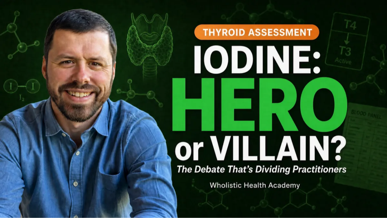
The Iodine Question: When More Helps, When More Harms, and Why It's Always About Pattern
By Michael Rutherford
Few nutrients in functional medicine split practitioner opinion as cleanly as iodine. One camp treats it as the missing key to most thyroid dysfunction. The other treats it as a third rail that flares Hashimoto's. The research supports a more nuanced position — and clinical pattern recognition is the bridge between the two camps.
Why Iodine Splits the Functional Medicine Conversation
Two practitioners can look at the same low-T4 lab result and reach opposite conclusions about iodine. One sees a textbook case of iodine deficiency and reaches for kelp or potassium iodide. The other sees an autoimmune red flag and warns the client to avoid iodine entirely. The frustrating reality is that neither practitioner is necessarily wrong — they're just looking at different patterns and treating them with universal rules instead of pattern-specific reasoning.
Iodine deficiency remains the most common cause of preventable hypothyroidism worldwide.1 At the same time, excess iodine intake — particularly in populations with adequate or replete baseline status — can trigger or accelerate autoimmune thyroid disease in susceptible individuals.1,2 Both statements are simultaneously true. The clinical question isn't "is iodine good or bad" but "where on the dose-response curve does this specific person sit, and what does the rest of their thyroid panel tell me about their susceptibility?"
The Physiology Refresher Most Iodine Debates Skip
The numerals in T4 and T3 aren't arbitrary — they count the iodine atoms attached to the tyrosine backbone.3 Thyroid hormone synthesis fundamentally cannot occur without iodine. Follicular cells of the thyroid actively concentrate iodine from the bloodstream through the sodium-iodide symporter, achieving intracellular concentrations 20-40 times higher than serum levels.
Once inside the follicular cell, thyroid peroxidase (TPO) — an enzyme that requires both iodine and iron as cofactors4 — oxidizes iodide and attaches it to tyrosine residues on thyroglobulin. Two iodinated tyrosines couple to form T4 (or, less commonly, T3 directly). The thyroid produces approximately 80% T4, with the remainder split between T3 and reverse T3.
Here is the part that matters for the iodine debate: TPO is also the primary autoantigen in Hashimoto's thyroiditis. The most commonly tested thyroid antibody — anti-TPO — targets this enzyme. When iodine intake rises sharply, the iodination process generates reactive oxygen species as a byproduct, increasing oxidative stress in thyroid tissue.5 In a person with no autoimmune predisposition and adequate antioxidant capacity, this is well-buffered. In a person with low selenium status, existing antibodies, or genetic susceptibility, that oxidative stress can drive antigen exposure and accelerate autoimmunity.1,2,5
Clinical Pearl: The enzyme that requires iodine to do its job is the same enzyme the immune system targets in autoimmune thyroiditis. This explains why iodine status is dose-dependent in a way most nutrients are not — and why dietary-level iodine and high-dose supplementation are functionally different interventions.
Ready to go deeper than this post can take you?
Mastering the Art of Functional Blood Chemistry is the in-depth practitioner course for pattern recognition and root cause clinical reasoning.
The Deficiency Pattern: First-World Iodine Inadequacy
In the developed world, severe iodine deficiency has become uncommon among populations that consume mainstream Western diets. But a specific subgroup is at meaningful risk: the health-conscious client who has restructured her diet around what she's been told is healthy.
The three reliable dietary iodine sources in a Western context are iodized table salt, conventional dairy (which picks up iodine from iodine-based sanitizers used in commercial milking operations), and seafood.1 Many of the dietary patterns popular in functional medicine — Paleo, autoimmune protocol, anti-inflammatory, dairy-free, "real salt only" — systematically eliminate all three sources.
Sea salts, Himalayan pink salt, and Redmond Real Salt have impressive trace mineral profiles and are nutritionally superior to refined iodized salt in many ways. But they contain negligible iodine. A client who has switched to "clean" salt, eliminated dairy, and rarely eats seafood has unintentionally eliminated her three best iodine sources without knowing it.
The classic deficiency lab pattern shows:
Pattern One — Non-Autoimmune Primary Hypothyroidism (Deficiency-Driven):
Elevated TSH · Low-normal free T4 · Low-normal free T3 · Negative TPO and thyroglobulin antibodies · Normal-to-low reverse T3 · Often accompanied by a dietary history consistent with iodine inadequacy
This is one of the patterns covered in detail in our comprehensive guide to thyroid panel interpretation. The clinical conclusion isn't aggressive iodine loading. It's modest dietary restoration — adding seafood, considering reintroduction of iodized salt in cooking, and supporting cofactors. Within three to six months, the panel typically normalizes if the cause was truly dietary.
The Excess Pattern: When Iodine Drives the Dysfunction
The second pattern is the opposite case — and it's the one that has made many functional medicine practitioners cautious about iodine over the past decade.
Two well-described mechanisms make excess iodine clinically problematic. The Wolff-Chaikoff effect describes the thyroid's protective response to acute iodine excess: a temporary downregulation of hormone synthesis that prevents thyrotoxicosis. In most people, the thyroid "escapes" this within days. In some — particularly those with underlying autoimmune disease or high baseline iodine status — escape fails and persistent hypothyroidism develops.1
The Jod-Basedow phenomenon is the opposite presentation: iodine-induced hyperthyroidism, typically in individuals with previously iodine-deficient nodular goiter who receive sudden iodine repletion.
But the most clinically relevant excess-iodine story in functional practice isn't acute thyrotoxicity. It's chronic supplementation driving autoimmune flare. Population-level data have consistently shown that introduction of universal salt iodization, while reducing endemic goiter, modestly increases population-level rates of autoimmune thyroiditis.1,2 At the individual level, high-dose iodine supplementation in someone with even subclinical autoimmunity can substantially increase antibody titers and accelerate gland destruction.
Pattern Two — Iodine-Triggered Autoimmunity:
Normal-to-elevated TSH · Normal or borderline-low free T4 · Low-normal free T3 · Rising TPO antibodies (often substantially elevated) · History of high-dose iodine supplementation or recent introduction of kelp/iodine-containing products
The intervention here is not more iodine. It is discontinuation, selenium repletion, and addressing the gut, stress, and infectious drivers of the underlying autoimmune process. Antibody titers typically trend downward over six to twelve months when iodine exposure is normalized.
The Cofactor Reality: Why Iodine Alone Is Almost Never the Answer
Even when iodine is genuinely low and supplementation is appropriate, iodine in isolation is rarely the right intervention. Thyroid hormone production and conversion require an interlocking nutrient system, and supplementing any single component without addressing the others tends to produce disappointing results at best and adverse outcomes at worst.
Selenium sits at the center of this system. The three deiodinase enzymes that convert T4 to active T3 (and inactivate hormone via reverse T3) are all selenium-dependent selenoproteins.3,6 Without adequate selenium, supplemental iodine has no efficient way to be activated into the form the body actually uses. Beyond conversion, selenium-containing glutathione peroxidases protect thyroid tissue from the oxidative byproducts of hormone synthesis — providing the antioxidant buffer that prevents iodine-driven inflammation from progressing to autoimmunity.5,7
The selenium-iodine interaction has been confirmed in multiple clinical contexts. Meta-analyses show selenium supplementation reduces TPO antibody titers in Hashimoto's thyroiditis, with the effect attributed both to reduced oxidative stress and to improved deiodinase function.7,8,9 This is why most experienced functional practitioners now consider selenium status the non-negotiable prerequisite before any iodine intervention.
The full nutrient picture includes:
›Iron — required as a cofactor for TPO; deficiency impairs hormone synthesis regardless of iodine availability4
›Zinc — supports TSH receptor function and is required for deiodinase activity3
›Copper — required for deiodinase enzyme function; deficiency impairs T4 to T3 conversion10
›Vitamin A — supports thyroid hormone receptor sensitivity3
›Tyrosine — the amino acid backbone iodine is attached to during hormone synthesis
The clinical implication is straightforward: an iodine intervention without baseline assessment of selenium and iron is taking on risk that's largely avoidable.
Pattern Recognition for the Iodine Question
This is where the three-pattern framework for thyroid interpretation cuts through the noise. Rather than asking "should this person take iodine," the more productive question is "which of the three patterns is dominant, and what does the antibody layer add to the picture?"
Primary hypothyroidism with negative antibodies and a dietary case for deficiency. This is the cleanest iodine-restoration scenario. Modest dietary repletion through seafood, conventional dairy reintroduction (if tolerated), or judicious use of iodized salt — combined with selenium support — typically resolves the picture without milligram-level supplementation.
Poor T4-to-T3 conversion (normal T4, low T3, often elevated reverse T3). This pattern is rarely about iodine. It's almost always about selenium, zinc, chronic stress, inflammation, or insulin resistance impairing deiodinase function. Adding iodine to a conversion problem doesn't help — and the reverse T3 elevation is a downstream signal of upstream problems, not a primary issue to target.
Stress-induced suppression (low or low-normal TSH with low T4/T3). This pattern reflects HPT axis suppression from chronic stress, severe caloric restriction, or systemic illness. Iodine doesn't address the upstream stress signal. Lifestyle, sleep, food intake, and adrenal support are the interventions.
Antibodies are the universal layer that gets evaluated separately from the dominant pattern. Positive TPO or thyroglobulin antibodies — at any titer meaningfully above the reference floor — should make any practitioner conservative with iodine, regardless of which pattern looks dominant. This is the cardinal rule that prevents the most common iodine-related clinical errors.
For a deeper exploration of how to integrate these patterns systematically across an entire blood chemistry panel, see our framework on clinical reasoning and pattern recognition in blood chemistry.
The Clinical Assessment Workflow
A practical workflow for the iodine question in a new client looks like this. First, run a complete thyroid panel — TSH, free T4, free T3, reverse T3, TPO antibodies, and thyroglobulin antibodies — alongside the broader chemistry that contextualizes thyroid function. A meaningful thyroid assessment isn't the thyroid markers in isolation. Blood sugar regulation, inflammatory markers, and iron status all shape thyroid output, and interpreting the panel without that wider context routinely produces the wrong intervention. Thyroid dysfunction is so frequently a downstream signal of upstream metabolic or immune disruption that reading TSH and free hormones in a vacuum is one of the most common clinical errors in the field. Second, take a detailed dietary and supplement history with specific attention to salt type, dairy intake, seafood frequency, and any current or recent iodine-containing supplements (including kelp, multivitamins, prenatals, and "thyroid support" formulas). Third, assess selenium and iron status before considering any iodine intervention. Fourth, classify the dominant pattern using the three-pattern framework, with antibodies as the separate layer.
The intervention plan follows directly from the pattern. The question is rarely "iodine or not" in isolation — it's "what does this entire pattern call for, and where does iodine fit, if at all."
Continue Your Education
Master the Art of Functional Blood Chemistry
The iodine question is one piece of a much larger pattern-recognition framework. Mastering the Art of Functional Blood Chemistry teaches the complete clinical reasoning system — integrating thyroid, adrenal, blood sugar, and immune patterns into a unified approach to root cause assessment.
Explore the CourseFor health coaches, NTPs, FDN-Ps, and nutrition professionals.
Frequently Asked Questions
How do I distinguish a true iodine deficiency from another cause of low thyroid hormones?
Three pieces of information together usually clarify the picture: the lab pattern (elevated TSH with low-normal free hormones and negative antibodies suggests deficiency rather than autoimmunity), the dietary history (a clear restriction of all three primary iodine sources), and the response to modest food-first restoration. Urinary iodine testing can add information but reflects recent intake more than tissue status, so it's best used as one data point rather than a standalone test.
Is iodized salt safe for clients with Hashimoto's thyroiditis?
Dietary-level iodine from iodized salt in cooking is generally well tolerated even in autoimmune thyroid disease. The concerning intervention is milligram-level supplementation — particularly iodine doses in the 1 mg to 50 mg range that have become popular in some functional protocols. Dietary iodine and supplemental iodine are functionally different interventions that should be evaluated separately.
Why does selenium status matter so much before iodine supplementation?
Selenium is the cofactor for every deiodinase enzyme that converts T4 to active T3.3,6 It also protects thyroid tissue from oxidative damage produced during hormone synthesis.5 Iodine supplementation without adequate selenium increases oxidative burden on the gland and reduces the ability to convert hormone — the combination most likely to drive autoimmune flares. Selenium repletion before iodine intervention is one of the most consistent findings in the functional thyroid literature.
What testing best establishes iodine status?
No single test is perfect. Spot urinary iodine reflects recent intake. 24-hour urinary iodine is more reliable but logistically demanding. Iodine loading tests have interpretive complexities and aren't universally accepted. In practice, a careful dietary history combined with a complete thyroid panel — particularly the antibody status — gives more clinically actionable information than any single iodine test in isolation.
Are there populations where iodine needs are different?
Yes. Pregnancy and lactation substantially increase iodine requirements — the developing fetus depends entirely on maternal thyroid hormone in the first trimester for brain development. Prenatal protocols should include adequate iodine (typically 150-220 mcg daily) alongside selenium and iron. This is one context where the deficiency risk significantly outweighs the autoimmunity risk for most individuals, though antibody status should still inform the approach.
References
- Zimmermann, M. B., & Boelaert, K. (2015). Iodine deficiency and thyroid disorders. The Lancet Diabetes & Endocrinology, 3(4), 286-295. https://doi.org/10.1016/S2213-8587(14)70225-6
- Triggiani, V., Tafaro, E., Giagulli, V. A., Sabbà, C., Resta, F., Licchelli, B., & Guastamacchia, E. (2009). Role of iodine, selenium and other micronutrients in thyroid function and disorders. Endocrine, Metabolic & Immune Disorders-Drug Targets, 9(3), 277-294.
- Chaker, L., Bianco, A. C., Jonklaas, J., & Peeters, R. P. (2017). Hypothyroidism. The Lancet, 390(10101), 1550-1562. https://doi.org/10.1016/S0140-6736(17)30703-1
- Beard, J. L., Borel, M. J., & Derr, J. (1990). Impaired thermoregulation and thyroid function in iron-deficiency anemia. American Journal of Clinical Nutrition, 52(5), 813-819.
- Ventura, M., Melo, M., & Carrilho, F. (2017). Selenium and thyroid disease: From pathophysiology to treatment. International Journal of Endocrinology, 2017, 1297658. https://doi.org/10.1155/2017/1297658
- Bianco, A. C., Salvatore, D., Gereben, B., Berry, M. J., & Larsen, P. R. (2002). Biochemistry, cellular and molecular biology, and physiological roles of the iodothyronine selenodeiodinases. Endocrine Reviews, 23(1), 38-89. https://doi.org/10.1210/edrv.23.1.0455
- Köhrle, J., Jakob, F., Contempré, B., & Dumont, J. E. (2005). Selenium, the thyroid, and the endocrine system. Endocrine Reviews, 26(7), 944-984. https://doi.org/10.1210/er.2001-0034
- Toulis, K. A., Anastasilakis, A. D., Tzellos, T. G., Goulis, D. G., & Kouvelas, D. (2010). Selenium supplementation in the treatment of Hashimoto's thyroiditis: A systematic review and a meta-analysis. Thyroid, 20(10), 1163-1173. https://doi.org/10.1089/thy.2009.0351
- Wang, F., Wang, Y., Wang, L., Wang, X., Sun, C., Xing, M., & Zhao, Y. (2023). Selenium and thyroid diseases. Frontiers in Endocrinology, 14, 1133000. https://doi.org/10.3389/fendo.2023.1133000
- Olin, K. L., Walter, R. M., & Keen, C. L. (1994). Copper deficiency affects selenoglutathione peroxidase and selenodeiodinase activities and antioxidant defense in weanling rats. American Journal of Clinical Nutrition, 59(3), 654-658.
