Iron Panel Interpretation Guide: Why Ferritin Alone Isn't Enough
Mar 02, 2026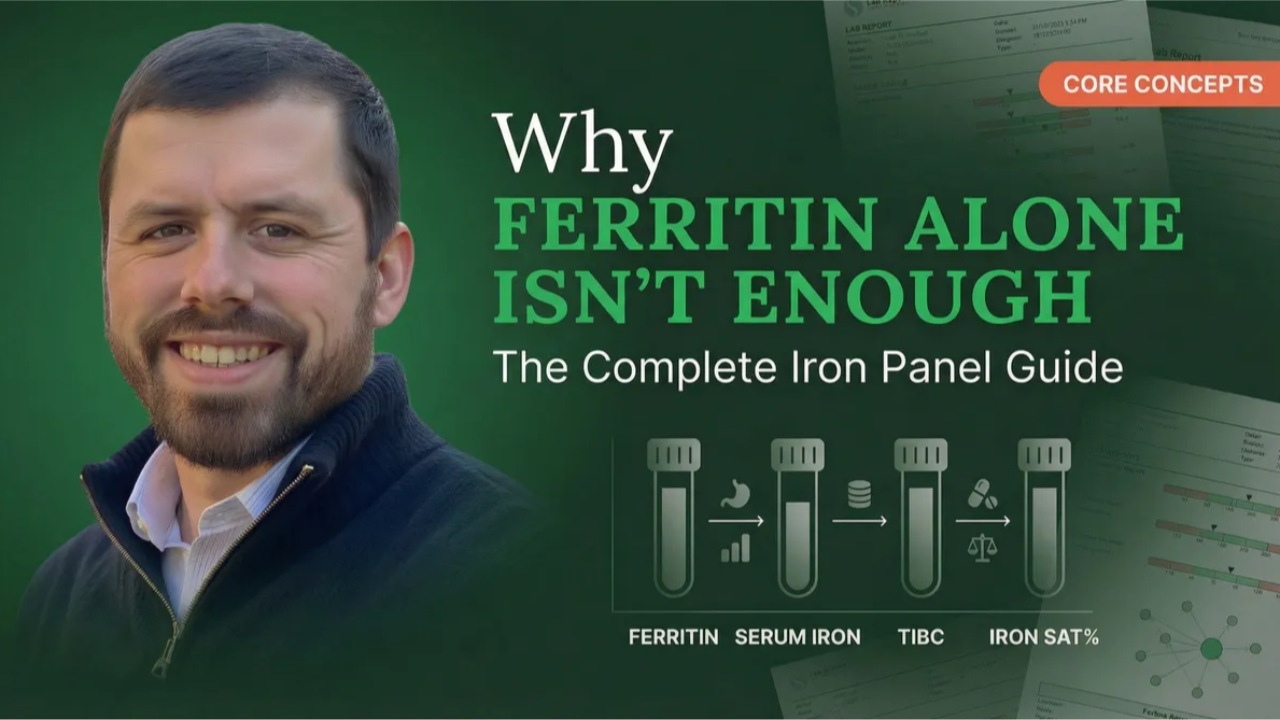
The Practitioner’s Guide to Iron Panel Interpretation: Why Ferritin Alone Isn’t Enough
Wholistic Health Academy • Iron Assessment • Clinical Education
Iron is the most paradoxical nutrient in human physiology—simultaneously essential for life and potentially toxic at elevated levels. It is the most damaging nutrient at both deficiency and excess, which makes accurate assessment critical and makes the common shortcut of relying on ferritin alone genuinely dangerous. A complete iron panel interpreted through pattern recognition is the only way to distinguish true iron deficiency from anemia of chronic disease, iron overload from inflammatory sequestration, and cofactor-driven dysfunction from actual iron insufficiency.
This guide covers the full iron panel, the four primary clinical patterns, the cofactor framework that changes how you approach iron status, and the referral criteria every practitioner should know. If you have been ordering ferritin and CBC alone, this will fundamentally change how you assess iron in your practice.
Why Is Ferritin Alone Misleading?
Ferritin serves a dual role: it is both an iron storage protein and an acute-phase reactant.1 This means ferritin rises with inflammation regardless of actual iron stores. A client with a ferritin of 90 ng/mL and elevated hs-CRP may actually be iron deficient—the inflammation is artificially inflating what appears to be adequate iron status. Conversely, a ferritin of 25 in a non-inflamed person genuinely reflects low iron stores.
This dual role has created a pervasive clinical error. Some practitioners recommend that ferritin should be maintained at 90 ng/mL or higher for everyone, citing studies from inflammatory bowel disease populations where inflammation masked true deficiency.2 But applying IBD thresholds to a general healthy population inappropriately elevates the target. The functional optimal range for ferritin is 30–125 ng/mL—and that number only means something when interpreted alongside the rest of the iron panel and the client’s inflammatory status.
One practical rule that cuts through this confusion: ferritin should rarely exceed serum iron (in US units). When ferritin is approaching or exceeding the serum iron value—for example, serum iron of 50 with ferritin of 45—that pattern suggests inflammation is involved, even if hs-CRP hasn’t crossed a conventional threshold.
What Does a Complete Iron Panel Include?
Iron status requires more markers than any other nutrient due to its complex regulation and multiple cofactor dependencies. A complete iron panel with ferritin is the bare minimum—yet most healthcare providers rely only on CBC and ferritin, which provides insufficient information for clinical decision-making.
The Complete Iron Panel — Functional Optimal Ranges
Ferritin: 30–125 ng/mL — Iron storage (also acute-phase reactant). Conventional range spans 12–300, which is far too wide to be clinically useful.
Serum Iron: 70–130 μg/dL — Circulating iron. Highly variable with dietary intake in the previous 24–48 hours. Fasting and avoiding supplements the day before testing improves reliability.
TIBC (Total Iron-Binding Capacity): 275–360 μg/dL — Measures the capacity of transferrin to bind iron. Moves in the opposite direction from iron status: high TIBC means the body is hungry for iron; low TIBC means the body is actively blocking iron absorption.
Iron Saturation: 25–40% — The ratio of serum iron to TIBC. Below 25% suggests deficiency; above 45% warrants investigation for overload.
Soluble Transferrin Receptor (sTfR): Lab-specific ranges — Critically, sTfR is not affected by inflammation, making it the most reliable marker for distinguishing true iron deficiency from anemia of chronic disease.3 The TfR:Ferritin Index further sharpens this differentiation.4
The key interpretive principle is that TIBC and serum iron should move in opposing directions. As iron increases, TIBC should decrease. When they move concordantly—both rising or both falling together—it suggests pathology rather than normal physiological variation.
What Are the Four Primary Iron Patterns?
Once you have the full panel, iron assessment becomes a pattern recognition exercise. Four primary patterns account for the vast majority of what you will see in practice. (For a broader overview of how pattern recognition works across all systems, see Pattern Recognition in Blood Chemistry: A Clinical Framework.)
Pattern 1: True Iron Deficiency — Low ferritin (<30), high TIBC, low iron saturation, elevated sTfR. The body is genuinely lacking iron and is increasing its binding capacity to capture whatever is available. This is the pattern where supplementation is appropriate—but cofactors should be addressed simultaneously.
Pattern 2: Anemia of Chronic Disease — Normal or elevated ferritin, normal or low TIBC, elevated CRP, low serum iron. This is the inflammatory trap: the body has adequate iron stores but is actively sequestering iron away from circulation through hepcidin-mediated mechanisms.5 When inflammatory cytokines like IL-6 rise, they stimulate hepcidin production, which blocks iron absorption from the gut and locks iron inside storage cells.6 Supplementing iron in this pattern is not only ineffective—it can increase oxidative stress.7 The correct intervention is to resolve the underlying inflammation first.
Pattern 3: Iron Overload — Elevated ferritin, high iron saturation, elevated serum iron. This pattern reflects iron accumulation beyond what the body can safely manage. Hereditary hemochromatosis is the most common genetic cause, typically confirmed through genetic testing and managed with therapeutic phlebotomy.8 Even emerging overload patterns—before markers reach overtly pathological levels—warrant attention, because iron overload drives ferroptosis, an iron-dependent form of cell death involving lipid peroxidation that contributes to liver damage, cardiovascular disease, and cancer risk.9 This is a pattern where early recognition and monitoring change outcomes.
Pattern 4: Iron Recycling Dysfunction — Variable ferritin (often low-normal), low serum iron (<70 μg/dL), normal to high TIBC, low iron saturation (<25%), often with low copper and low ceruloplasmin. This pattern looks like iron deficiency on the surface—but supplementing iron makes things worse because the problem is not iron supply. It is impaired iron recycling. Ceruloplasmin, a copper-dependent enzyme, is essential for converting stored iron into a usable form through its ferroxidase activity. When copper is deficient—whether from inadequate intake, zinc excess suppressing copper absorption, or vitamin A deficiency reducing ceruloplasmin production11—iron gets stuck. The root causes here are copper deficiency (primary or secondary), zinc excess, vitamin A deficiency, chronic oxidative stress, and in rare cases genetic ceruloplasmin variants. Addressing the copper-vitamin A axis is the intervention, not iron.
Why Do Cofactors Matter More Than Iron Itself?
This is the clinical insight that changes everything about iron assessment: the majority of functional iron deficiency cases involve cofactor deficiencies rather than true iron insufficiency. Eight essential cofactors are required for iron metabolism—copper, vitamin A, folate, B12, magnesium, vitamin D, B6, and zinc. When any of these are insufficient, iron cannot be properly absorbed, transported, stored, or incorporated into hemoglobin, regardless of how much iron is present or supplemented.
Vitamin A deficiency, for example, impairs iron mobilization from storage and contributes to functional anemia even when iron stores are adequate.10 It also reduces ceruloplasmin activity, which disrupts copper metabolism—and copper is itself essential for iron recycling through ferroxidase activity.11 These interconnections explain why clients who supplement iron for months without improvement often see resolution once cofactors are addressed.
Clinical Pearl: Beef Liver as the Iron Cofactor Solution
Beef liver’s effectiveness for iron status stems not primarily from its iron content, but from its comprehensive cofactor profile: copper, vitamin A, folate, B12, zinc, B6, vitamin C, and magnesium—all in one food source. For iron deficiency without anemia, desiccated beef liver supplementation addresses the cofactor picture simultaneously. For iron deficiency with anemia, beef liver combined with beef spleen provides a more comprehensive intervention than isolated iron supplementation in most cases.
This cofactor-first approach aligns with the broader clinical methodology taught in Mastering the Art of Functional Blood Chemistry: identify the root cause before reaching for the obvious intervention. In iron assessment, the root cause is often not iron at all.
How Does Menstrual Cycle Affect Iron Interpretation?
For menstruating clients, testing timing matters. A serum iron of 65 μg/dL on day 6–7 of the cycle (just after menstruation) may be acceptable since stores have just been drawn down. The same value on day 28 would be more concerning because stores should have recovered by that point in the cycle. Average menstrual loss is 50–70 mL per cycle, but some women lose 400+ mL, creating severe and recurrent iron depletion that no supplement protocol can overcome without addressing the underlying cause of heavy bleeding—whether hormonal imbalance, fibroids, or endometriosis.
Women with genetic hemochromatosis present a unique early pattern: menstruation may prevent the typical iron accumulation, keeping ferritin artificially low and masking the condition until menopause, when iron stores begin rising without the monthly offset. This is why a comprehensive iron panel matters at every stage—not just when overt deficiency or overload is suspected.
How Do You Apply This in Practice?
Iron assessment exemplifies why functional blood chemistry is a pattern recognition discipline, not a single-marker exercise. Every iron marker tells part of the story. Together, they reveal whether the problem is deficiency, inflammation, overload, or recycling dysfunction—and the intervention for each is fundamentally different. Tools like Blood Chem Studio organize the full iron panel alongside inflammatory markers and RBC indices, making these pattern distinctions visible at a glance. For the complete clinical education framework covering iron and every other major system, explore The Complete Guide to Functional Blood Chemistry Interpretation.
Master Iron Assessment and Every Other System
Mastering the Art of Functional Blood Chemistry covers the full iron panel, cofactor frameworks, pattern recognition, and clinical reasoning across all major systems.
Explore the CourseFrequently Asked Questions
What is the optimal ferritin level for women?
The functional optimal ferritin range is 30–125 ng/mL for both men and women. This is not gender-specific—what matters is interpreting ferritin alongside the complete iron panel and inflammatory markers. A ferritin of 40 in a non-inflamed person represents adequate stores, while a ferritin of 90 with elevated CRP may mask underlying deficiency.
Can you have iron deficiency with normal ferritin?
Yes. Because ferritin is an acute-phase reactant, inflammation can elevate ferritin even when true iron stores are depleted. This is why a complete iron panel—including serum iron, TIBC, iron saturation, and ideally sTfR—is essential. Soluble transferrin receptor is unaffected by inflammation and rises early in true tissue iron deficiency, making it the most reliable differentiator.3
Why does iron supplementation sometimes not work?
The three most common reasons are cofactor deficiencies (copper, vitamin A, B6, and zinc are required for iron utilization), inflammation-driven sequestration (hepcidin blocks absorption regardless of how much iron you take), and poor absorption from inadequate gastric acid. Addressing these root causes resolves the majority of non-responsive iron supplementation cases.
What is the difference between iron deficiency and anemia?
They are distinct conditions that can exist independently. You can have iron deficiency without anemia (depleted stores but hemoglobin still maintained), anemia without iron deficiency (B12, folate, or chronic disease-driven), iron deficiency anemia (both present), or even high iron with concurrent anemia. This is why relying on CBC alone to assess iron status leads to incomplete or incorrect conclusions.
What does high TIBC mean on a blood test?
High TIBC means the body is increasing its iron-binding capacity because it needs more iron. Think of it as the body creating more “trucks” to capture and transport iron because supply is low. High TIBC alongside low ferritin and low iron saturation is the classic true iron deficiency pattern. Conversely, low TIBC means the body is restricting its ability to absorb iron—typically because inflammation has triggered hepcidin to block iron uptake as a protective mechanism.
References
- Knovich, M. A., Storey, J. A., Coffman, L. G., Torti, S. V., & Torti, F. M. (2009). Ferritin for the clinician. Blood Reviews, 23(3), 95–104.
- Dignass, A. U., Gasche, C., Bettenworth, D., et al. (2015). European consensus on the diagnosis and management of iron deficiency and anaemia in inflammatory bowel diseases. Journal of Crohn’s and Colitis, 9(3), 211–222.
- Skikne, B. S., Punnonen, K., Caldron, P. H., et al. (2011). Improved differential diagnosis of anemia of chronic disease and iron deficiency anemia: a prospective multicenter evaluation of soluble transferrin receptor and the sTfR/log ferritin index. American Journal of Hematology, 86(11), 923–927.
- Punnonen, K., Irjala, K., & Rajamäki, A. (1997). Serum transferrin receptor and its ratio to serum ferritin in the diagnosis of iron deficiency. Blood, 89(3), 1052–1057.
- Weiss, G., & Goodnough, L. T. (2005). Anemia of chronic disease. New England Journal of Medicine, 352(10), 1011–1023.
- Nemeth, E., Rivera, S., Gabayan, V., et al. (2004). IL-6 mediates hypoferremia of inflammation by inducing the synthesis of the iron regulatory hormone hepcidin. Journal of Clinical Investigation, 113(9), 1271–1276.
- Lund, E. K., Wharf, S. G., Fairweather-Tait, S. J., & Johnson, I. T. (1999). Oral ferrous sulfate supplements increase the free radical-generating capacity of feces from healthy volunteers. American Journal of Clinical Nutrition, 69(2), 250–255.
- Bacon, B. R., Adams, P. C., Kowdley, K. V., Powell, L. W., & Tavill, A. S. (2011). Diagnosis and management of hemochromatosis: 2011 practice guideline by the AASLD. Hepatology, 54(1), 328–343.
- Dixon, S. J., Lemberg, K. M., Lamprecht, M. R., et al. (2012). Ferroptosis: an iron-dependent form of nonapoptotic cell death. Cell, 149(5), 1060–1072.
- Semba, R. D., & Bloem, M. W. (2002). The anemia of vitamin A deficiency: epidemiology and pathogenesis. European Journal of Clinical Nutrition, 56(4), 271–281.
- Smith, J. E., Ciaccio, E. I., Teng, J. I., & Goodman, D. S. (1976). The effects of vitamin A deficiency on the plasma concentration of ceruloplasmin. Journal of Laboratory and Clinical Medicine, 88(3), 427–434.
Michael Rutherford • Wholistic Health Academy • wholistichealthacademy.org
